Never had a full eye exam before? Here’s exactly what happens, step by step.
An eye examination checks two things: your eyeglass prescription and your eye health. It’s done by an optometrist or ophthalmologist, and it usually follows a fixed sequence of tests. Not every test applies to every patient — your eye care practitioner decides what’s needed based on your history and symptoms.
Here’s what a typical eye exam includes.
1. Medical and Eye History
Your exam starts with questions, not equipment.
Expect to be asked about:
- Past or current eye problems
- Medications you’re taking
- Family history of eye disease
- Health conditions like diabetes or hypertension
This matters more than it seems. Conditions like diabetes affect the blood vessels in your retina, so an accurate history often decides whether you’ll need a retina exam later. Answer honestly — everything else in the exam builds on this.
2. Vision Check (Visual Acuity Test)
Next, your eyesight is measured using a vision chart — one eye at a time, with the other covered.
- Charts vary by age, language, and literacy — including picture charts for children and non-readers
- If you wear glasses or contacts, bring them; your vision is checked with your current prescription too
- Lost or damaged glasses? Mention it before the test
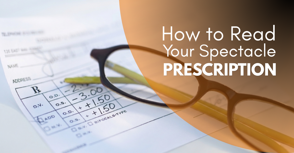
3. Refraction (Determining Your Prescription)
This is where your eyeglass prescription is calculated.
Even with normal vision, the practitioner checks your refractive status using an auto-refractor or retinoscope. This gives a starting number, which is then fine-tuned by testing different lens strengths against the vision chart until your eyes see clearest.
Lenses tested may be spherical, cylindrical, or both. This step can take several minutes — precision matters more than speed here.
4. Cycloplegic Eye Drops (Mainly for Children)
For children and young adults, drops may be used to dilate the pupil and temporarily relax the eye’s focusing muscles. This gives a more accurate prescription than refraction alone.
5. Eye Muscle Coordination Test
You’ll be asked to track a moving light or object. This checks the strength and coordination of your eye muscles.
- Weakness or misalignment (squint) may require vision therapy — a structured set of eye exercises
- Some squints need surgery instead; not all respond to therapy alone
6. Torchlight Examination
A quick check using a pen torch to spot obvious external abnormalities and test pupil response. It’s often used when a full slit-lamp exam isn’t possible — for example, with very young children.
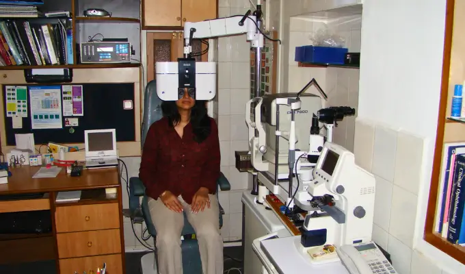
7. Slit-Lamp Examination
Your eyes are examined under a microscope with a bright light source (the slit lamp), covering everything from eyelids and lashes to the internal lens. Special dye may be used to check for tear film problems or eye surface damage.
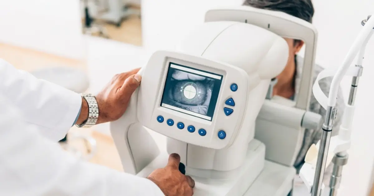
8. Eye Pressure Test (Glaucoma Screening)
This measures your intraocular pressure using a tonometer — either a puff of air or a gentle probe (with numbing drops).
High eye pressure can silently damage the optic nerve and cause permanent vision loss before you notice symptoms. That’s why glaucoma is often called “the silent thief of vision.” If your pressure is elevated, expect follow-up tests or a referral to a glaucoma specialist.
9. Colour Vision Test
You’ll view cards with colored dot patterns. Difficulty spotting the numbers or shapes may indicate colour blindness.
10. Fundus Examination (Retina Check)
The final step: examining the back of your eye — the retina, blood vessels, optic disc, and choroid — using ophthalmoscopy. Dilating drops give a clearer view but cause temporary blurry vision and light sensitivity afterward.
A fundus exam is essential if you have:
- Diabetes or high blood pressure
- Family history of glaucoma, macular degeneration, or retinal disease
- Sudden or recent vision loss
- High myopia (severe nearsightedness)
- Flashes of light or floating black spots in your vision
What Happens After the Eye Exam?
Your practitioner reviews all findings and explains them to you. If you need glasses, a prescription is issued. If anything needs closer attention, you may be scheduled for a follow-up — for contact lens fitting, detailed refraction, binocular vision assessment, or a dilated fundus exam.